Skip to content

SurgiPrintยฎ


Leading The Edge In High-Definition 3D Surgical Models.

What we do ? Illustrated application - how 3D model helps Surgical planning

Useful application

Surgical Planning and operation guidance

3D models transform surgical planning by providing unparalleled precision and depth, enabling surgeons to visualize and strategize complex procedures with greater accuracy and confidence.

Education - University

3D models provide students with a detailed, interactive representation of human anatomy, allowing for a deeper understanding of surgical techniques, anatomical variations, and procedural planning.

Robotic Training

3D models serve as an invaluable training tool for robotic surgery, offering a realistic and interactive environment for surgeons to hone their skills and master intricate robotic maneuvers.

The process: We make doctors lives' easy

Clinics can upload the DICOM and receive the 3D model and the required measurements in less than 3 days.

1. Imaging

CT Scans, PET Scans, MRI Medical images are uploaded to our secured server in seconds.

2. From 2D to 3D

Our AI algorithm, along with our team of dedicated radiologists will transform the traditional 2D images into a digital 3D model.

surgiprint 3D medical image

3. Easy link access

SurgiPrint will send a link by email which is immediately accessible on any device just by a click. No requirement of any software installation.

Our Main expertise: HPB - Hepato-Pancreato-Biliary
The Power of AI in Tumor Detection

๐Ÿš€ Revolutionizing Liver Health: The Power of AI in Tumor Detection ๐Ÿš€

In the quest for advancing medical care, Artificial Intelligence (AI) has taken a monumental leap in transforming liver health diagnostics. AI-driven tumor detection in the liver presents a myriad of benefits that span across the healthcare spectrum, from hospitals and patients to doctors and healthcare insurers. Hereโ€™s why:

๐Ÿฅ For Hospitals: Implementing AI for liver tumor detection streamlines diagnostic processes, significantly reducing the time between screening and diagnosis. This efficiency not only enhances patient throughput but also optimizes the utilization of medical resources, paving the way for higher standards of care and operational excellence.

๐Ÿ‘ฉโ€๐Ÿ”ฌ For Patients: Early and accurate detection is paramount in battling liver tumors. AI algorithms offer unparalleled precision, catching tumors at their nascent stages when treatment options are more effective and less invasive. This early intervention can drastically improve patient outcomes, offering hope and a chance for a healthier future.

๐Ÿฉบ For Doctors: AI tools act as an invaluable second opinion, providing doctors with insights derived from analyzing vast datasets that the human eye might overlook. This support empowers physicians with a higher degree of confidence in their diagnostic decisions, enabling them to tailor treatment plans that are precise, personalized, and effective.

๐Ÿ’ณ For Healthcare Insurance: Early detection and accurate diagnosis lead to cost-effective treatments with better outcomes, reducing the financial strain on both insurers and insured. By investing in AI technologies, healthcare insurers can mitigate high treatment costs associated with advanced liver diseases, thus lowering premiums and enhancing coverage for patients.

In conclusion, AI in liver tumor detection is not just an innovation; it’s a transformative force that benefits all stakeholders in the healthcare ecosystem. As we continue to harness the potential of AI, the future of liver health and overall healthcare looks brighter than ever.

The different anatomy we can reconstruct

Surgical planning Benefits Explained

๐Ÿš€ Revolutionizing Medical Imaging with 3D Model Reconstruction ๐Ÿš€

In today’s rapidly evolving medical field, the leap from traditional 2D CT scans and MRIs to dynamic 3D model reconstructions marks a monumental shift in patient care and surgical precision. ๐Ÿฅ๐Ÿ’ก

Why is this transition so pivotal? Let’s dive in:

  1. Enhanced Visualization ๐ŸŒˆ: 3D models provide an unprecedented view of the human body’s intricate structures, allowing doctors and surgeons to navigate and understand complex anatomies with greater depth and clarity.

  2. Precision Planning ๐Ÿ”: Pre-surgical planning has never been more accurate. With 3D reconstructions, surgeons can strategize procedures with pinpoint accuracy, reducing uncertainties and improving outcomes.

  3. Customized Patient Care ๐Ÿ‘ค: Every patient is unique, and 3D modeling embraces this, offering tailored visualizations based on individual scans. This personalized approach ensures better preparation and patient-specific treatment strategies.

  4. Educational Excellence ๐Ÿ“š: For medical students and professionals alike, 3D models serve as powerful educational tools, enhancing learning and training experiences by providing hands-on, interactive studies of human anatomy and surgical techniques.

  5. Innovative Solutions ๐ŸŒŸ: The field of medical research and device development benefits greatly from 3D models, paving the way for the design of cutting-edge medical devices and therapeutic techniques.

In conclusion, the shift to 3D model reconstruction from traditional imaging methods is not just a technological upgrade; it’s a paradigm shift in medical diagnostics, treatment planning, and education. The future of healthcare is here, and it’s three-dimensional. ๐ŸŒ๐Ÿ”ฌ

Patient Education

Having the surgery explained well to a patient comfort his trust in both the doctor and the surgical operation. 3D reconstructed anatomy models are proving invaluable in facilitating patient understanding of prospective surgeries and procedures.

Pre-Op

The surgeon will have a significantly improved understanding of the work he has to do. Surgeons can now gain the benefit of an unobstructed 3D patient-specific view of hidden tissues and blood vessels.

During Op

Studies show the 3 following improvements : duration of surgical operations is decreased significantly ; as well as the average volume of bleeding ; drastic increase of the number of lymph node dissections.

Post-Op

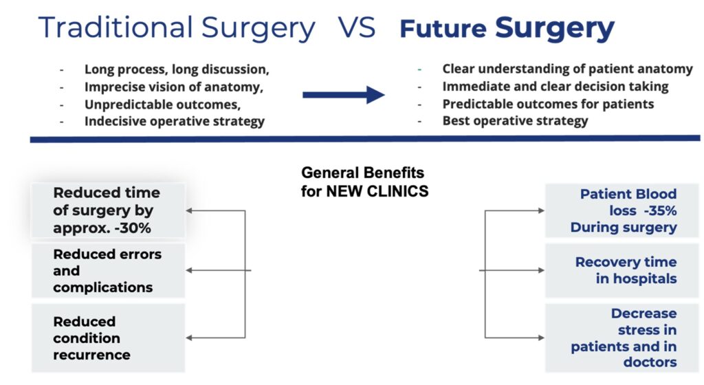
The overall outcome of the surgery prove to have much improved results by the following metrics : Decrease of the Postoperative flatus recovery time ; Decrease of the Duration of hospitalization. The studies show a general satisfaction rate improvement for both the patient and the doctor of over 30%.